Technology

Discover scalable, instrument-free single cell sequencing technology from Parse Bioscience

Technology Overview


Resources

Explore our collection of resources to learn more about technology and its applications from leading researchers

Resources Overview

Company

Providing researchers single cell sequencing with unprecedented scale and ease

About Parse

BLOG › Best Practices › Getting Started with scRNA-seq: Experimental Design and Sample Preparation

Getting Started with scRNA-seq: Experimental Design and Sample Preparation

October 21, 2024
|
12 min read
Updated:October 21, 2024


Still wondering about the benefits of adding single cell RNA sequencing (scRNA-seq) to your research? A quick scan of some peer-reviewed literature can answer this question, and help give you guidance on how to implement scRNA-seq in your lab. 

ScRNA-seq provides invaluable insights into the unique transcriptional profiles of individual cells within tissues or organs. For example, examining a tumor at single cell resolution allows researchers to explore how each cell type contributes to the tumor’s function and its surrounding microenvironment. This can play a critical role in the development of targeted therapies.

Another powerful application of scRNA-seq is identifying rare cell types. For instance, this study in a mouse model of Alzheimer’s Disease found that C5aR1 antagonism suppresses inflammatory glial responses and alters cellular signaling. By combining single cell and single nucleus RNA-seq analysis of hippocampi, the study identified C5aR1-dependent neurotoxic, disease-associated microglia clusters and provided insights into communication among hippocampal cell subtypes.

Moreover, scRNA-seq helps identify co-regulatory gene networks and gene modules, offering a higher-resolution picture of cellular processes on a transcript-by-transcript basis. 

While the benefits of adding scRNA-seq may be becoming clearer, mastering any new technology comes with somewhat of a learning curve, and scRNA-seq is no different.

This article will provide an overview of the applications and challenges of scRNA-seq experimental design, along with practical tips and resources to prepare a single cell or nuclei suspension from various sample types. 

There are two main areas to consider when approaching scRNA-seq experiment for the first time:

Important Considerations for an Experimental Design

Designing a successful scRNA-seq experiment involves several key considerations, from sample selection and preparation to quality control. There are a few critical factors to consider when planning a scRNA-seq experiment to optimize the experiment while avoiding common pitfalls.

Sample Size

An important consideration that could even impact the choice of scRNA-seq technology is the sample size — researchers need to sequence enough cells to answer their biological question. 

While microfluidic-based technologies may limit the ease of scalable sample size, combinatorial barcoding technologies don’t have that limitation as they are plate-based.

Experimental planning tools are ideal for making such decisions. Numerous tools like the Single Cell Experimental Planner enable scientists to plan experiments based on specific needs. Important points to consider are:

  • What type of study — is it for a publication, a pilot, a small research project, a cell atlas, a drug screening?
  • Number of cells needed/available
  • Number of samples available
  • Sample type
  • Sequencing needs and suggested platforms

Sample Type

Before planning the experiment, a decision point is choosing between sequencing whole cells or just nuclei. Each approach has its pros and cons, and the decision depends on the research question and nature of the samples. Choosing nuclei sequencing is beneficial for cells difficult to dissociate without compromising viability.

For instance, highly fibrous tissues — brain, skin, tumors with a high amount of extracellular matrix — are challenging to dissociate, often resulting in cell death.
Moreover, some samples, like cells from three-dimensional cultures, present challenges. Once extracted from their three-dimensional matrix, these cells perish quickly.

For such scenarios, the alternative is single nuclei sequencing. Most genes reside in the nucleus, so sequencing nuclei captures nearly the same transcriptomic data despite a nominal loss of RNA from the cytosol.

In clinical contexts, nuclei are invaluable. They permit immediate freezing of tissue samples, whether sourced from the operating room or from sizable tissue harvests.
Opting for nuclei over whole cells can also facilitate extraction from samples stored in liquid nitrogen.

Moreover, the constraints of the chosen technology play a role in deciding between cells and nuclei. If a researcher deals with larger cells and the chosen technology restricts cell size, as seen with pico-wells or certain droplet-based systems, then nuclei become the inevitable choice.

Number of Samples

The number of samples analyzed plays a crucial role as it affects the accuracy of overall results and increases the power of statistical tests. One reason for manuscript rejection is the need for more technical or biological replicates within the study. The same holds true for studies that leverage scRNA-seq as a primary research method.

But if the sample size is inadequate, pooling becomes a viable solution. There might be a need for more cells for the assay, especially with embryonic samples, rare organisms, or when viable cells are scarce. In such cases, combining samples can address the issue.

For instance, pooling several samples from distinct model organisms (like drosophila embryos) or combining multiple sections of an identical tissue creates enough biological mass to meet minimum cell counts of snRNA-Seq sample preparation. Additionally, the ability to fix samples allows the accumulation of cells or nuclei over time, making pooling logistically more feasible.
Replication is another key aspect of good experimental design that involves the use of an adequate number of samples.

There is a difference between technical and biological replicates and their respective applications (Figure 1).

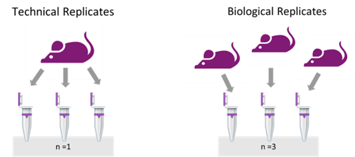
Figure 1: A well-thought experimental design considers technical and biological replicates to account for sources of variability.

A technical replicate measures the noise of the protocols or equipment by dividing the same sample into two or three sub-samples and processing them separately.

In contrast, biological replication entails examining biologically different samples under identical conditions — multiple subjects with the same disease or cells from different donors treated with the same stimulus. This approach captures the inherent variability in biological systems and verifies the experiment’s reproducibility.

Bioinformatic Support

A common concern for the novice scRNA-seq researcher is bioinformatics support. The computational aspects of single cell sequencing can be daunting. The good news is that there are now many user-friendly software options available that don’t require extensive programming knowledge, so that bench scientists now don’t have to double as computer scientists to obtain reliable and publication ready data.

It is also good practice consulting with bioinformaticians within the department or core facility during the planning phase of your experiment.

Fresh or Fixed?

One last critical decision is the use of fresh or fixed samples.

When running an experiment, researchers are focused on capturing a snapshot in biology, whether it be a specific developmental stage, a series of individual time points within a time course experiment.

Cellular metabolism and gene expression rapidly change once cells are removed from their physiological environment. Consequently, delays in sample processing lead to results that reflect stress responses rather than true biological states. Therefore, minimizing processing time is crucial in scRNA-seq experiments.

Fixation addresses the issue as researchers can dissociate the tissue, fix it, and store it.

Storing fixed samples in the freezer for later use streamlines many experimental logistics and lab research situations, such as:

Clinical Laboratory Setting

Working with fresh samples in a clinical laboratory presents several logistical challenges. For instance, tissues arriving from the operating room can do so at unpredictable times, disrupting experiment schedules and staffing. Even with lab prepared tissues, where timing is known, there’s the added task of booking instruments and arranging transport to the core facility.

Large scale projects

Large-scale projects can be particularly demanding in scRNA-seq. Time course experiments require sequential sample collection over long periods. Processing fresh samples for each time point can lead to batch effects that obscure the study variables. With fixation, this is no longer an obstacle.

Experiment setup time is another significant concern. Sample preparation from tissues and organs is labor-intensive, maintaining cell viability throughout the process is crucial, requiring careful coordination among lab members and efficient resource allocation. Planning sample accrual and fixation puts researchers back in charge of their own experiments.

Technical Variability

Another critical aspect is managing technical variability. Samples collected over extended periods are susceptible to batch effects due to inconsistencies in collection times, environmental factors, and even technician performance. These variabilities, often hard to control, can significantly impact experimental results.

Using a plate-based combinatorial barcoding method can be a valuable solution, as the user can fix, store, and later run up to 96 samples with a single kit.

Fixing cells or nuclei post-preparation offers flexibility to conduct experiments at a more convenient time (Table 1).

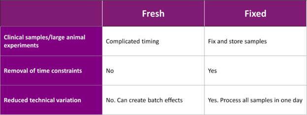
Table 1: Common considerations of scRNA-Seq experimental design: comparison of fixed and freshly prepared cells.

Sample Preparation

An informative dataset requires viable cells or nuclei to begin with. The ideal sample viability should be between 70% and 90%, with intact cell morphology. To do so, a sample preparation method needs to minimize temperature effects, reduce cell clumps and debris, and ensure accurate cell counts.

Single Cell or Nuclei Suspension Methods

Creating a single cell suspension can be done in several ways, depending on the tissue. Density gradient, vortexing plant nuclei into solution, gentle pipetting of cells mixed with enzymes for organoid suspensions, or using commercially available enzyme cocktails are all viable options.

Each tissue type has its own specific recipe for dissociation, so it’s important to refer to protocols from the literature, such as those on protocols.io. These protocols often provide detailed instructions, including enzyme types, mixing steps, and times required.

Whenever possible, sample manipulation should be gentle and fast. For example, density centrifugation — using Ficoll or Optiprep — is a simple yet effective technique for separating viable cells from debris. It works well for PBMC fractionation, excluding dead cells and even cleaning up nuclei. If dissociating mouse brain tissue, density centrifugation helps remove myelin sheath, which, if left in the sample, causes aggregation and introduces noise into the sequencing data.

For those new to tissue dissociation, it is good practice to start with guides such as the Worthington Tissue Dissociation Guide, which provides detailed protocols for different tissue types, including enzyme concentrations and mixing steps. Another useful resource is Miltenyi Biotec, a company that offers enzyme cocktails and plug-and-play kits for generating single-cell suspensions.

Automated tissue dissociation instruments are very effective methods to obtain high quality suspensions. Both the Miltenyi’s gentleMACS™ Dissociator, and the S2 Genomics’s Singulator™ Platform, ensure rapid and reproducible solid tissue dissociation.

Temperature Control

Maintaining a stable temperature is vital for scRNA-Seq sample preparation.

When extracting cells, maintaining a cold environment helps arrest their metabolic functions. For example, peripheral blood mononuclear cells (PBMCs) held at 4°C for three hours still appear viable, while those held at room temperature for the same duration start to die, extrude cellular contents, and clump together. This degradation impacts the quality of the library and sequencing data (Figure 2).

Figure 2: Know the sample temperature that cells require to maintain viability and arrest metabolic functions.

Once the suspension is created, cells should be placed immediately on ice to cool and halt metabolic activity. This reduces the upregulation of stress response genes, which can skew the data.

Avoid Debris

A viable single cell suspension has minimal cell clumping and debris.

Why does aggregation happen? Aggregation typically stems from dead cells, tissue debris, or the presence of cations like calcium and magnesium in the media, which act as magnets for dead cells. Over-pelleting cells during centrifugation can also cause clumping.

These issues are easy to address. The user can filter the debris out, use media without calcium or magnesium (such as HEPES or Hanks’ buffered salt), and test different centrifugation speeds and durations to avoid over-pelleting (Figure 3).

Figure 3: Single cell/nuclei suspension should have minimal debris and aggregation (<5%).

Sample QC: Cell Count and Cell Viability

Accurate cell counting is critical before proceeding, as it ensures a successful experiment. The parameters used to assess the suspension—uniform single cells, minimal clumps, minimal debris, and accurate cell counts—are key to producing high-quality data.

It is important to observe the cells under a microscope to ensure high viability. For example, fibroblasts in a healthy state are bright, with clear nuclei. After treatment or stress, their membranes may crack, and cytosol leaks out, indicating cell death. This visual assessment is easy and cost-effective using a wide-field microscope with a 20x lens.

When working with nuclei, assessing nuclear quality by microscopy is equally important. During apoptosis, chromatin begins to break down, causing nuclei to enlarge, and eventually, apoptotic bodies form and exit the nuclear membrane. These changes are observable with DAPI (4′,6-diamidino-2-phenylindole) or Hoechst staining. Good nuclei should be intact and uniformly stained, with no signs of broken chromatin. Apoptotic nuclei appear larger and deformed (Figure 4).

Figure 4: DAPI Staining shows nuclear degradation. Degradation appears as nuclear membrane blebbing, chromatin degradation, and release of apoptotic bodies.

Conclusions

Like mastering any other art, creating the best conditions for any successful and highly informative experiment requires some trials and errors.

Proper design and planning are fundamental to create such conditions, but they are often overlooked aspects of scientific research. ScRNA-Seq experiments are no exception.

Though securing a high-quality cell suspension is the most challenging aspect, adequate preparation can simplify it and be the difference between bad data and exceptional data.

About the Author

Laura Tabellini Pierre

Laura Tabellini Pierre, MSc, is a scientific and technical writer at Parse Biosciences with extensive experience in immunology, encompassing both academic and R&D research.